


Dr. Leila Soudah is a specialist obstetrician on healthcare of women and their children during pregnancy, childbirth and post natal care. During pregnancy division of stages are measured in trimester from the day of the last menstrual period. Each trimester, Dr. Leila Soudah Clinic offers a comprehensive step to ensure a safe and healthy pregnancy journey both the mother and the baby.
- Ultrasound:
- First Trimester Screening:
- A combination of Nuchal translucency, Ultrasound Examination procedure by well-trained licensed sonographer which measures the collection of fluid under the skin behind the fetal neck. NT thickness could be increase for fetus with chromosomal abnormalities, cardiac defects and other genetic syndromes. And correlation with the maternal serum free Beta HCG and PAPP-A provides effective method of early screening for trisomy 13, 18, 21. The combination of Nuchal translucency and maternal serum free Beta HCG and PAPP-A improves the detection to 90%. There is now evidence that the detection rate can increase to about 95% and the false positive rate can be reduced to 2.5 % by also examining the following markers:
- Nasal Bone: These are two oblong bones, placed side by side at the middle and upper part of the face and form by their junction " the bridge" of the nose. For fetuses with trisomy 21 and other chromosomal abnormalities the nasal bone is hypo plastic or not visible at 11-13weeks of gestation.
- Facial Angle: It is the wide angle in a line drawn over the palate and between the maxilla and the forehead. Recent studies suggest that fetuses with trisomy 21 have a flat profile because the maxilla (upper jaw) is small and set back.
- Tricuspid Valve: Flow of blood between right atrium to right ventricle. This is one of the markers that contribute in the screening for both major cardiac defects.
- DuctusVenosus: blood flow of the umbilical vein directly to the fetal heart. It is a part of fetal circulation. Presence of normal flow will reduce the risk for trisomy 21 and cardiac abnormalities and the presence of reversed A- wave will increase the risk.
- Uterine Artery PI: It is the two arteries both left and right that supplies blood to the uterus in females. In correlation with maternal history, blood pressure is measured in a certain way both arms to calculate the risk for developing Pre- eclampsia, hypertension is detected.
- Second Trimester Screening:
- Integrated Sequential screening
- Alpha-fetoprotein (AFP): A blood test that measures the level of AFP in the mother's blood during pregnancy. AFP is a protein normally produces by the fetal liver and is present in the fluid surrounding the fetus (amniotic fluid) and crosses the placenta into the mother's blood.
- HCG: Human chorionic gonadotropin hormone produced by the placenta.
- Free Estriol: Hormone produced by the placenta.
- Inhibin A: Hormone produced by the placenta.
- Amniocentesis
- Anomaly or detailed scan
- Third Trimester Screening:
- Group B streptococcus screening
- Doppler Ultrasound/ Doppler Study
- Cardiotocography (CTG)
- Fetal Echocardiography
- Fetal Growth Scan
- Fetal Fibronectin
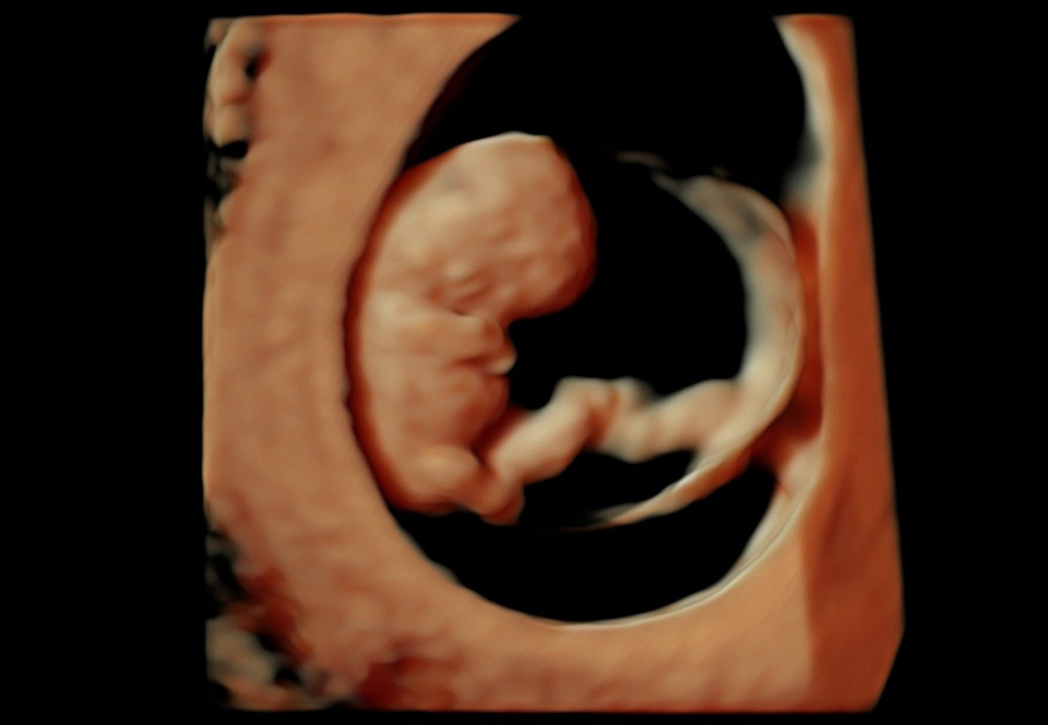
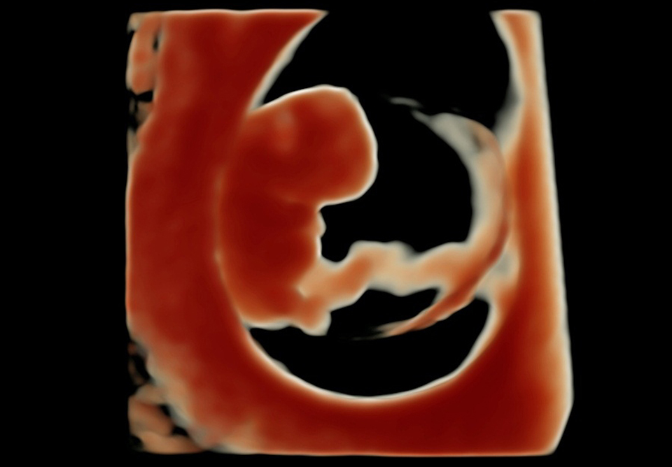
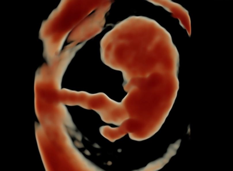
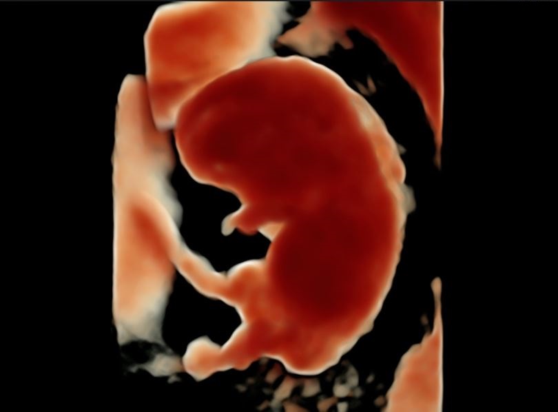
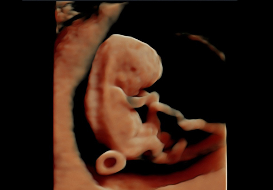
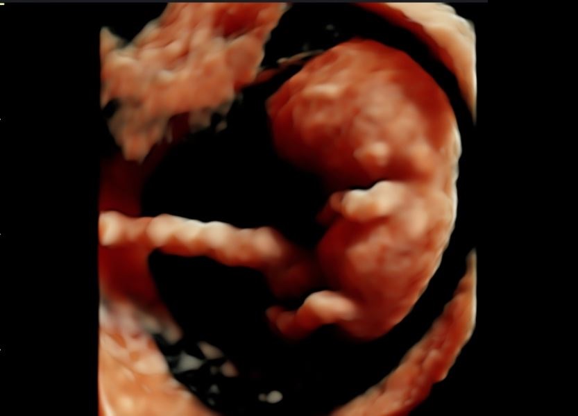
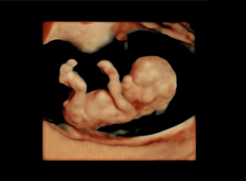
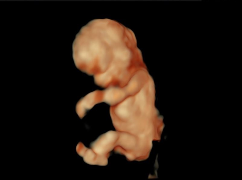
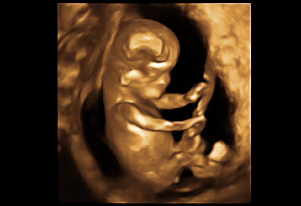
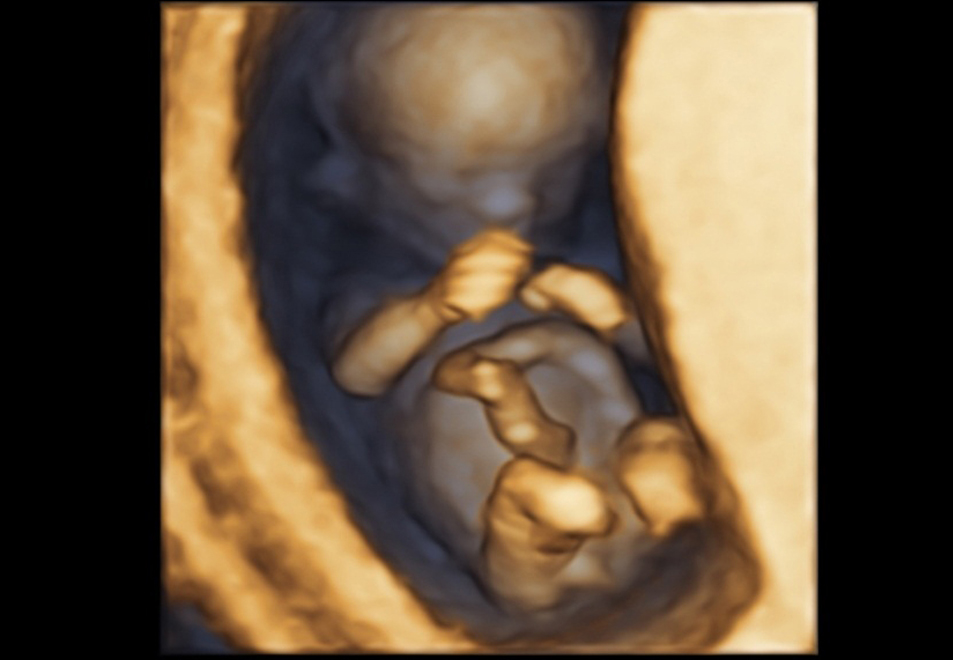
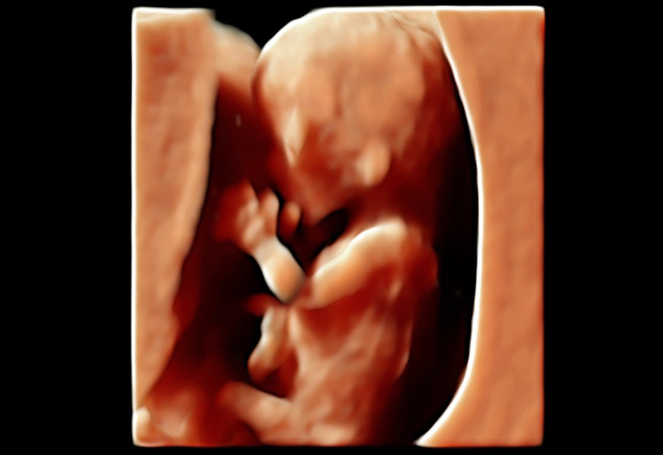
2D/3D/4D and the Latest High Definition Live Ultrasound - every detail from the inside of the woman's reproductive system and baby's details during pregnancy can be seen clearly using this ultrasound.


It is a painless one hour in-clinic Ultrasound procedure and easy screening test that a pregnant woman can do and go back to her normal routine after the procedure. This test will rule out birth defect like neural tube, chromosomal abnormality such as trisomy 13, 18, 21. Every pregnant patient should be offered this kind of test regardless of her age. Dr. Leila Soudah has been a licensed sonographer both under England and Germany Fetal Medicine Foundation since 2004 to perform this test and calculate the risk of abnormality for your baby and listed as one of the sonographer with good audit results under FMF England.
This test is done between 11+0 to 13+6 weeks of pregnancy.


If the first trimester screening is abnormal, integrated sequential test can be offered, chorionic villus sampling and amniocentesis depending on the high risk calculation of the patient.
During this stage, typically you will see the Doctor once every four weeks unless you have a condition or complications that call for more frequent visits for checkups. In here, blood tests will be done, such as the glucose screening test to check for gestational diabetes. In addition, in cases first trimester screening shows high risk fully integrated screening is offered. Second trimester screening includes the following:


This screening test is performed by taking blood sample of the mother between 14+0 to 19+6 weeks of pregnancy. This includes the following blood parameter to measure:
When a pregnant woman has both first trimester (NT and PAPP -A) and second trimester (AFP, HCG, Free Estriol ,Inhibin A) screening test performed, the ability if the test to detect an abnormality is greater than using just one screening independently, which is having 96% detection rate. Most all cases of Down syndrome can be detested when both 1st and 2nd trimester are used.
Patient who missed first trimester screening entirely, on her second trimester, Quadruple test can be performed on the mother's serum and this test has 81% sensitivity of detection for Down syndrome.
It is an invasive medical procedure used in prenatal diagnosis for chromosomal abnormalities and fetal infections, in which a small amount of amniotic fluid that contains the fetal tissue is aspirated in the amniotic sac surrounding the fetus under the guide of the Ultrasound.
This is a one hour ultrasound procedure wherein Dr. Leila Soudah takes a precise and closer look to examine each part of the fetal body, determine the position of the placenta, assess the amount of amniotic fluid, and measure fetal growth. Special attention is paid to the vital developing organ such as brain, face, spine, heart, stomach, bowel, kidneys and limb.
At this point, through the high definition ultrasound imaging technology, Dr. Leila a licensed and experienced sonographer will be able to pinpoint multiple potential problems that can arise in developing fetuses. Although not all possible physical conditions can be picked up by the scan, a significant proportion of major irregularities can be found and necessary precaution needs to be taken and right care will be advised along the pregnancy.
Undergoing this procedure the Doctor will tell you about everything that she sees, unless you advise that there are certain things that you don't want to know, such as the baby's gender.
Further, if serious problems turn out upon the outcome of the scan, Dr. Leila Soudah gives and supports the opportunity to discuss all possible options and appropriate medical attention and suitable decision.
The third trimester is the best time for evaluating fetal growth, the quantity of amniotic fluid as well as the vessels of the mother and the baby, thus recognizing in time if the placenta functions properly or if there are any dangers for the baby. Especially in cases of abnormal second trimester screening tests, multiple pregnancy, a low level of amniotic fluid and the risk of insufficient supply to the placenta.
A series of screening test are done at Dr. Leila Soudah Clinic to safeguard the preparation for childbirth.


Vaginal and rectal swabs are taken at 35 to 37 weeks of pregnancy to detect group B strep bacteria. Although group B strep can be present in up to 30% of all healthy women, it's the leading cause of life-threatening infections in newborns and can also cause mental retardation, impaired vision, and hearing loss. Women who test positive are treated in pattern presented with the lab results during delivery to protect the baby from acquiring the infection at birth.
A procedure used to check the blood flow through the fetus's umbilical cord. Here at Dr. Leila Soudah Clinic Color Doppler is use as standard ultrasound methods to produce a picture of a blood vessel. Also, a computer converts the Doppler sounds into colors that are overlaid on the image of the blood vessel and that represent the speed and direction of blood flow through the vessel. Power Doppler is a special type of color Doppler. This kind of test is compulsory to pregnant patients with history of previous abortion, small babies or premature deliveries. Doppler Ultrasound gives the good information if the baby is getting enough blood supply.
This is done starting from 28 weeks of pregnancy for assessing fetal well-being monitoring and recording the fetal heartbeat and uterine contractions to prevent adverse fetal outcome such as pre-mature delivery.
It is an ultrasound that requires special Doctor Skills to examine the heart shape, size, position, blood flow and function. In addition, this helps to rule out cardiac anomaly where information guides in assessing the right Hospital with the good Neonatal care unit and the mode of delivery.
The purpose of this scan is to check that the baby is growing well and that the pregnancy is developing normally. Further this scan particularly important if you have had pregnancy complications or problems in a previous pregnancy.
The growth of the baby will be assessed by measuring the head, abdomen and femur length (thigh bone). This way the Doctor can estimate the baby's weight and give you an idea as to how big it will be at birth. The appearance and position of the placenta is also checked, and the amniotic fluid around the baby is measured.
Importance of this scan as well, is you have the idea of the mode of delivery which enables you to prepare yourself and the coming of the baby.
Fetal Fibronectin in cervicovaginal secretion used as an aid to rapidly assess the risk of preterm delivery. This is a painless procedure where Dr. Leila collects from the pregnant patient cervix by a special transport swab.
This test is more accurate in combination with the cervix length assessment (measuring the cervix length). Interpretation of positive result, an increased risk for preterm delivery is highly occurrence thus the Doctor gives appropriate advices and medication to prevent the preterm labor and subsequently delivery.